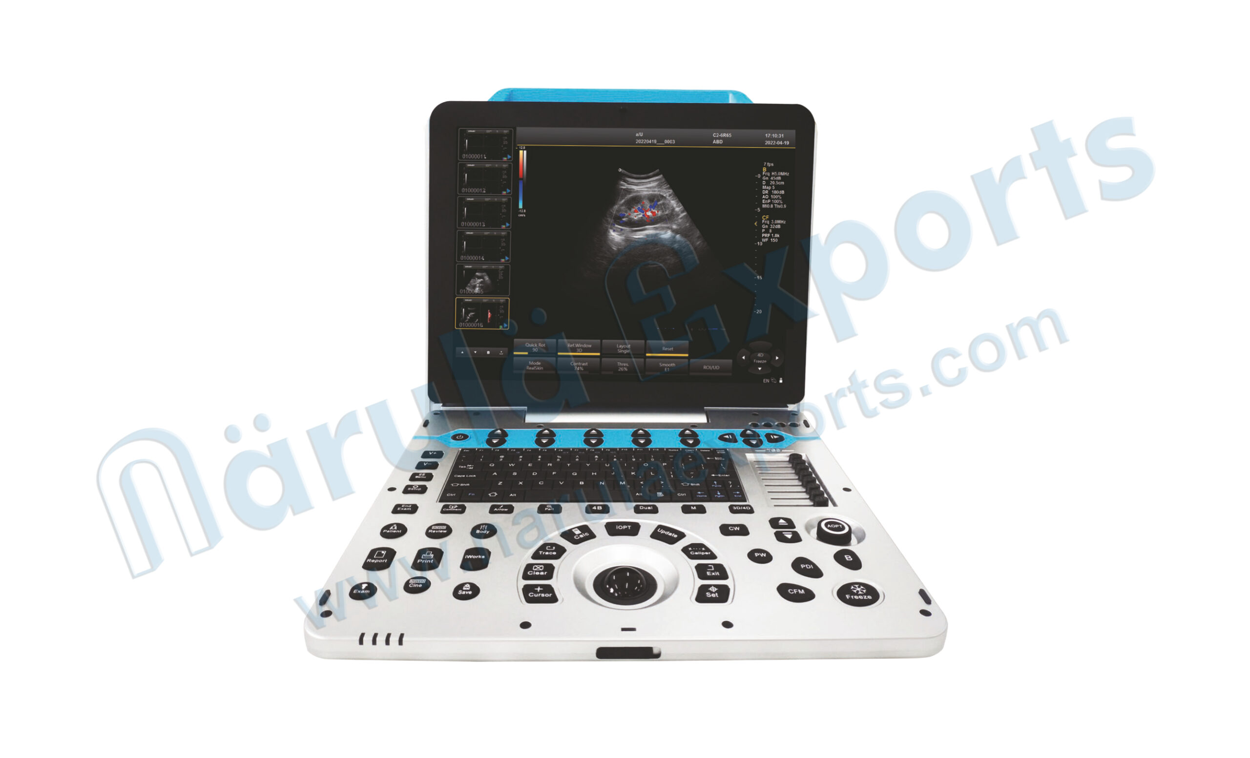


Model No.: 84-4080
Ultrasound that you can afford
Equipped with a high-resolution medical display, adopts multi-beam parallel technology and sub-array element transducers, and its superior image clarity perfectly meets the needs of women’s health care.
Smooth Workflow
Easy operation process, quick response to diagnostic needs, can easily deal with complex situations in mobile diagnosis.
Features
- High resolution medical 15.6 inch display.
- The display is adjustable from 0 to 30°.
- Hard disk dynamic and static image storage, real-time sharing.
- Spectrum envelope function.
- Built-in large capacity lithium battery (detachable).
Brilliant Ergonomics
- High resolution 15.6″LED with tilting functionality.
- User-friendly keyboard and controls.
Excellent Image Quality
- Tissue Harmonic lmaging (THl) enhancing contrast resolution.
- Quick image optimization by IP (lmage Processing).
- 8-segment TGC allowing delicate image adjustment.
Powerful Workflow
One-Key-Save, user-defined keys bringing great efficiency to your daily diagnosis.
Clinical Versatility
7 probes with abundant measurement packages covering traditional ultrasound applications and emerging fields such as urology, MSK and anesthesia.
Weight
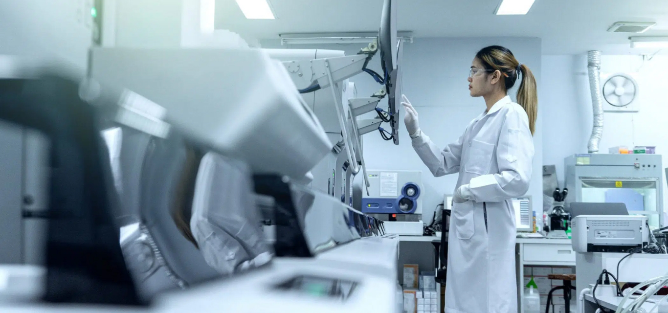
Only 9.0kg. Take it to your patients.
Cost
Diagnosis needs and budget conflict? Have a try on portable medical ultrasound system.
The multi-purpose user presets, comprehensive measurement & report system, built-in easy view image archive system, quick image storage/retrieve/transfer, one-button direct print, make the complete workflow better than what you can dream of.
Micron Imaging Technology
Micron imaging technology, real-time tracking of specific signals at the edges of different tissues, to achieve edge enhancement, and monitor each pixel at the same time, optimize the internal signal of the organization and perfectly integrate the edge information and the internal pixel information of the organization to restore the real and delicate, excellent level contrast Two-dimensional image.

Harmonic Imaging Technology (THI)
It improves image clarity by improving tissue contrast resolution, spatial resolution, and eliminating near-field artifacts. It is mainly used for the diagnosis of cardiovascular and abdominal diseases. It plays an important role in evaluating the lesion area and boundary division of patients with imaging difficulties. The technology has been fully approved by clinicians.
Harmonic technology retains the second harmonic signal to the greatest extent based on removing the fundamental signal, which increases the signal strength by more than 30% compared with the traditional signal processing, reduces noise and artifacts, and improves the contrast resolution of tissue images.

Trapezoid Imaging
Trapezoid imaging is a kind of expanded imaging, which is transformed into a trapezoid based on the original rectangle, and the left and right sides are expanded to a certain extent, achieving a wider field of view. The principle of ultrasound imaging is to scan the human body with ultrasonic sound beams, and obtain images of internal organs by receiving and processing the reflected signals.

Automatic Spectrum Tracking Measurement Technology
Ultrasound Doppler technology is used in the ultrasound system for examining the heart and arteries and veins. It is necessary to extract relevant parameters from the Doppler spectrogram to evaluate the hemodynamic status of the heart and blood vessels. The disadvantage of manual detection is that the operator’s marking of the peak velocity is relatively monotonous and time-consuming, with poor repeatability and low estimation accuracy, and during the detection, in order to mark the peak velocity, the operator needs to interrupt the acquisition of Doppler signals, which makes it impossible to estimate in real time.
This host contains an automatic envelope detection module, which can automatically track the time-related changes of the peak blood flow velocity and average velocity, and display them in real time on the Doppler spectrogram.

Clinical Image Cases

Part of the Probe Display

Sub-Array Technology
The dedicated high-density probe adopts a brand-new array design technology and a unique sub-array element technology. The second cutting of independent wafers can completely control the entire process of wafer vibration, thereby reducing side lobe artifacts and enhancing the fine resolution of tissues. The boundary between adjacent strong echo reflectors is sharper. It fully demonstrates the high-resolution images brought by the high-density probe, perfectly presents the image details, and increases the accuracy of clinical diagnosis.