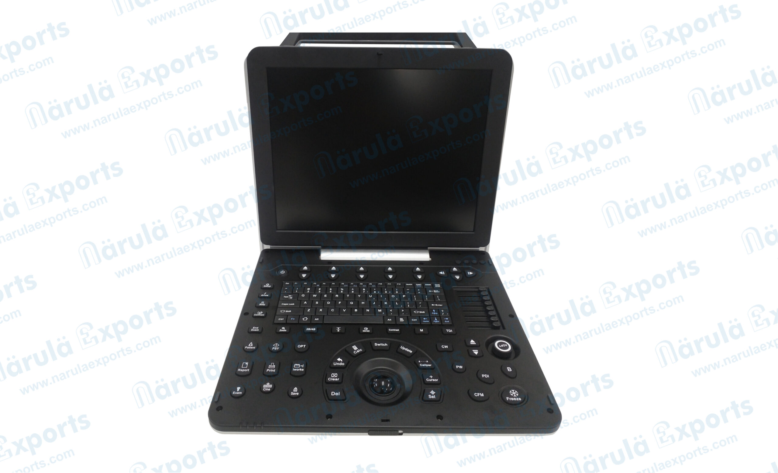


Model No.: 84-4010
Based on advanced design concepts and technological innovation, it R&D team developed this color doppler ultrasonic diagnostic instrument.
The intelligent operating system, abundant software measurement packages, convenient and quick measurement methods and humanized design, improve the efficiency and accuracy of doctors’ diagnosis.
- More Accurate
- Smarter
- More Real
- Compactor
- Faster
Features
- 15-Inch full-view medical HD display on the main screen.
- USB 3.0 interface clinical picture, video, report storage, export.
- Real-time wide-field imaging (WFOV).
- Trapezoidal imaging, continuous Doppler imaging.
- Free anatomy 3M imaging.
- IMT automatic measurement of blood vessel intima.
- Free arm 3D imaging, real time 4D imaging.
- Spatial composite imaging, speckle noise removal technology.
- Tissue Doppler Imaging (TDI).
Portable
Smaller size, easy to move, suitable for ultrasound examination applications in various environments, long standby time.
More powerful
To have powerful image processing functions, a wide range of technical applications, harmonic imaging, harmonic fusion, free arm 3D, elastography, trapezoidal imaging. contrast imaging.
Wide range of applications
The systemic application type is mainly used in the diagnosis and research of abdominal and obstetric and gynecological ultrasound. It has the application capabilities of cardiovascular peripheral blood vessels, obstetrics and gynecology, abdomen, fetal heart, superficial tissues and small organs, intraluminal and puncture interventional ultrasound with powerful 4D analysis functions.
Light As Swallow Accurate And Comprehensive
- Dual probe socket, portable color Doppler ultrasound system.
- Using a new computing engine and image processing technology.
- Has a sensitive blood flow detection capability and a wide range of probe adaptation capabilities.
- Meet the abdomen, obstetrics and gynecology, heart, small organs, superficial blood vessels, musculoskeletal, four-dimensional, etc.
- Except routine examination needs, can also meet the clinical needs of other specialized clinics.

b/sri: real-time dual image display
M mode
Used to check the tissue movement and display the direction of a sound beam selected by the user over time.

Trapezoidal Imaging
Refers to converting the line data of a linear array probe into a trapezoidal image through coordinate transformation and interpolation, which is an extended imaging.

Freehand Elastography Mode
Freehand elastography can help doctors distinguish between soft/hard lesions and surrounding tissues.

Wide-Field Imaging
Also known as ultra-wide-field imaging Compared with ordinary ultrasound imaging wide-field imaging provides a new perspective for clinical diagnosis, and has a very important clinical significance for observing large lesions and the relationship between the lesions and surrounding tissues. Diagnostic significance.

Contrast Imaging
The use of significantly different echo characteristics from human soft tissue, or acoustic characteristic impedance (i.e. specific acoustic impedance)

High Definition And High Quality
Freehand 3D Imaging Mode
Provides a method for generating 3D images when using standard linear array, convex array, and cavity probe inspection. The process of freehand 3D Imaging is to obtain a series of frame Images (refer to the above figure, move the probe in parallel at a uniform speed) apply volume rendering technology to reconstruct the volume data and display the 3D rendered image.

Volume 3D/4D Imaging Mode
4D Imaging, also known as real-time 3D imaging, provides an interactive means of viewing dynamic 3D imaging. The freehand 3D imaging mode has different probe movement speeds, during 4D imaging inspection, the volume probe is fixed at one location and cannot be moved. The machinery inside the probe the component can perform stable continuous scanning of different positions by swinging, so as to obtain a series of continuous and stable frame Images. It can be seen that the quality of the 4D rendered image is significantly higher than the 3D imaging with the bare hand.

Gaining Style And Combining Strengths Interoperability
Gaining style and combining strengths intaroperability, with the advent of the 5G era and the era of big data, Interconnectivity, remote consultation, etc. Will be the future development trend of medical equipment diversified external interfaces, and the dicom 3.0 protocol.
Anatomical M-mode
Has only one m-sampling line, which has limitations for moving examination tissues. especially for difficult patients. The anatomical M mode makes up for the lack of traditional M-mode for the examination of patients with difficult imaging, and it provides multiple M-sampling lines. To enable you to perform more effective motion analysis on M mode Images at different angles and positions.

Puncture Enhancement
Automatic detection of the needle body automatic deflection of the sound beam, and smart puncture enhancement technology make the puncture display in the human body more intuitive.

Real-time dual-frame display allows simultaneous activate both modes

Tissue Doppler imaging mode
Abbreviated as TDI mode, using Doppler principle to estimate tissue motion, such as the speed of myocardial motion. The TDI mode can obtain motion information and generate color-coded images of tissue motion speed.

Color M Mode
Abbreviated as MC mode, used for cardiac examination applications. Color blood flow uses speed and variance color maps to make colors superimposed on M-mode images. Color blood flow covers B-mode images and M-mode timeline.

D Mode
Also known as PW mode. PW Doppler allows you to selectively check blood flow data from a small area (i.e. the sampling volume) PW Doppler displays blood flow information through a constantly moving spectrum, visually Describes the functional relationship between blood flow velocity and time.

Automatic IMT Measurement
The thickness of the intima of the blood vessel is an important indicator for predicting the risk of cardiovascular disease. The automatic measurement technique of the intima can automatically measure the thickness of the intima in the near and far fields of the blood vessel and automatically optimize the measurement angle.

Clinical Images Some Cases Show