


Model No.: 84-4075
- Abdomen
- Urology
- Small Parts
- Thopedics
And More
The machine has been designed from the relentless focus on delivering uncompromising performance at a cost-effective price Equipped with high-end imaging technology, color images more delicate, higher clarity. With ergonomic design, High resolution medical display, image loss free.
Smart Collaboration Reliable Operation
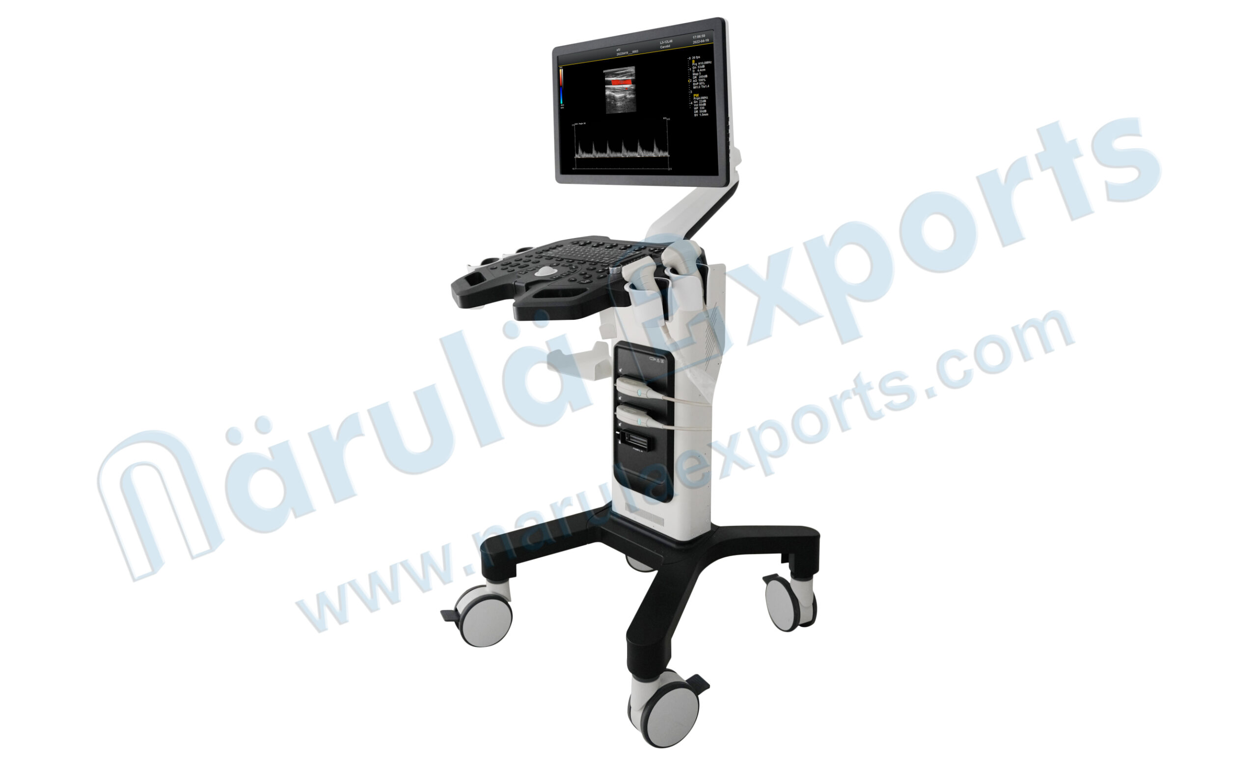
- Home screen 21.5 inch medical HD display.
- Adjustable monitor stand.
- Trackball: for easy operation.
- Detachable probe hanger.
- All-in-one keyboard for easy operation.
- Fully Activated Interoperable Three Probe Interface.
- Flexibly rotated casters with orientation lock.
Smooth Workflow
One-click intelligent optimization, fast access to quality images All-in-one clipboard Smooth processing Edge enhancement processing The host built-in
Excellent Image Quality
Spectral pulse Doppler Directional energy Doppler Spatial composite imaging Tissue harmonic imaging technique 4B imaging mode
User-Friendly Operation
Backlit, easy-to-use control panel the classical ergonomic design With DICOM3.0 protocol, PACS system can be connected
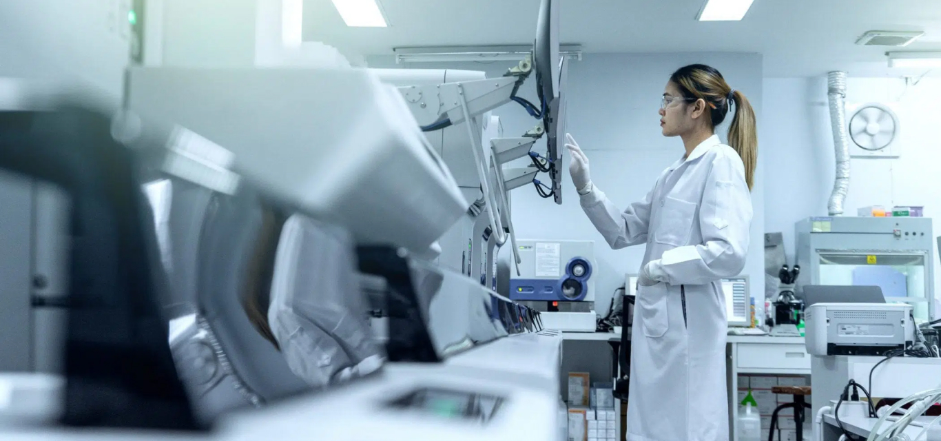
As an intelligent application of color Doppler ultrasound, it adopts the independent research and development and innovative ultrasonic operation platform, which is equipped with an excellent-performance hardware structure and a rapid information transmission module, combining advanced probe configurations and technologies. Compared with the former ultrasound imaging system, it presents a clearer, more realistic, more sensitive and smoother ultrasound image. With Various imaging function, perfect clinical solutions, this machine operates simpler and more efficiently, which considerably improve staff efficiency and optimize interactions experience.
Clear Image Visualization
The research and development has spent three years, integrating the most advanced design concept and technological innovation, to create full digital high performance full digital color Doppler ultrasound diagnostic instrument.
Intelligent operation process, humanized appearance design and intimate human-computer interaction as a whole, so that doctors in the process of clinical diagnosis will focus on the patient itself.

Micron Imaging Technology
Micron imaging technology, real-time tracking of specific signals at the edges of different tissues, to achieve edge enhancement, and monitor each pixel at the same time, optimize the internal signal of the organization and perfectly integrate the edge information and the internal pixel information of the organization to restore the real and delicate, excellent level contrast Two-dimensional image.
Tissue harmonic imaging (THI)
By improving tissue contrast resolution, spatial resolution and eliminating near-field artifact, image clarity can be improved. It is mainly used in the diagnosis of cardiovascular and abdominal diseases and plays an important role in the evaluation of lesion areas and demarcation of difficult imaging. This technology has been fully recognized by clinicians.
Harmonic technology retains the second harmonic signal to the maximum extent on the basis of removing the fundamental signal, which increases the signal intensity by more than 30% compared with the traditional signal processing, reduces noise and artifacts, and improves the contrast resolution, of tissue image.

Trapezoid Imaging
Trapezoid imaging is a kind of expanded imaging, which is transformed into a trapezoid based on the original rectangle, and the left and right sides are expanded to a certain extent, achieving a wider field of view. The principle of ultrasound imaging is to scan the human body with ultrasonic sound beams, and obtain images of internal organs by receiving and processing the reflected signals.

Carotid Spectrum
Spectral ultrasonography of carotid artery can provide a noninvasive, simple and reproducible method for the diagnosis of atherosclerosis. However, multi-parameter analysis should be advocated in the analysis of detection results, Besides the flow velocity of relevant vascular segments, pulsing index, spectral morphology, blood flow direction and blood flow sound should also be considered.
Carotid ultrasound is helpful to determine the nature of the ischemic cerebrovascular disease of carotid artery atheromatous plaque and stability, and to determine the degree of carotid atherosclerosis and carotid stenosis, especially in the display has the advantages on the change of the arterial wall structure, for the early prevention and treatment of atherosclerosis provide objective basis, actively treating atherosclerosis and carotid stenosis in preventing ischemic brain have important significance.

HD Liver Imaging Effect
2D real-time ultrasound imaging is mainly used for the change of liver morphology. Ultrasound examination shows the pathological image of liver, which belongs to the change of acoustic physical properties. For the same lesion, different stages of disease development, Ultrasonic image performance is different.


Excellent Image Convenient Operation
Various software measurement packages to meet the increasingly complex clinical examination needs; intelligent operation process, simple user interface & plenty imaging technology that canQ significantly improve the daily work efficiency, help doctors quickly collect images, and achieve more accurate output of diagnostic results.
Imaging with clarity and accuracy
Transducers